Research

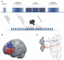
Transcranial magnetic stimulation (TMS) is an FDA-approved therapy for patients with major depressive disorder for whom several psychopharmacologic treatments have failed to produce clinical benefits. In this pilot study, we studied a brief three-day, personalized TMS protocol based on each individual’s functional connectivity map obtained from resting state fMRI data (i.e., an fMRI-guided protocol). We showed that the magnitude of the subgenual anterior cingulate’s response to short bursts of transcranial magnetic stimulation (single pulses of TMS) delivered to an fMRI-guided cortical target was associated with the degree of depression symptom improvement over the course of the three-day TMS treatment which was also delivered to the fMRI-guided cortical target. These results suggest the subgenual anterior cingulate’s acute response to single pulses of TMS may be a useful biomarker for the development of more effective, personalized TMS treatments in the future. This paper was recognized as one of the 2023 Leading Research Achievements by Brain and Behavior Research Foundation Grantees, Prize Winners, and Scientific Council Members.

A key factor in designing randomized clinical trials is the sample size required to achieve a particular level of power to detect the benefit of a treatment. Sample size calculations depend upon the expected benefits of a treatment (effect size), the accuracy of measurement of the primary outcome, and the level of power specified by the investigators. In this study, we show that radiomic models, which leverage complex brain MRI patterns and machine learning, can be utilized in clinical trials with protocols that incorporate baseline MR imaging to significantly increase statistical power to detect treatment effects. Akin to the historical control paradigm, we propose to utilize a radiomic prediction model to generate a pseudo-control sample for each individual in the trial of interest. Because the variability of expected outcome across patients can mask our ability to detect treatment effects, we can increase the power to detect a treatment effect in a clinical trial by reducing that variability through using radiomic predictors as surrogates.

The presence of a paramagnetic rim around a white matter lesion has recently been shown to be a hallmark of a particular pathological type of multiple sclerosis lesion. Increased prevalence of these paramagnetic rim lesions is associated with a more severe disease course in MS, but manual identification is time-consuming. We present APRL, a method to automatically detect PRLs on 3T T2*-phase images.

To acquire larger samples for answering complex questions in neuroscience, researchers have increasingly turned to multi-site neuroimaging studies. However, these studies are hindered by differences in images acquired across multiple sites. Contemporaneously with the increase in popularity of multi-center imaging, the use of machine learning (ML) in neuroimaging has also become commonplace. In our recently accepted paper in Human Brain Mapping, we demonstrate that methods for removing site effects in mean and variance may not be sufficient for ML. This stems from the fact that such methods fail to address how correlations between measurements can vary across sites. Data from the Alzheimer's Disease Neuroimaging Initiative is used to show that considerable differences in covariance exist across sites and that popular harmonization techniques do not address this issue. We then propose a novel harmonization method called Correcting Covariance Batch Effects (CovBat) that removes site effects in mean, variance, and covariance. We apply CovBat and show that within-site correlation matrices are successfully harmonized. Furthermore, we find that ML methods are unable to distinguish scanner manufacturer after our proposed harmonization is applied, and that the CovBat-harmonized data retain accurate prediction of disease group. 

While many findings in neuroimaging studies pertain to multiple imaging modalities, statistical methods underlying intermodal comparisons have varied. In our recently published paper in Human Brain Mapping, we propose the Simple Permutation-based Intermodal CorrEspondence (SPICE) test--an intuitive but powerful method for testing correspondence between two imaging modalities. The SPICE test differs from prior related methods in that it requires no assumptions about the brain’s complex spatial structure and leverages subject-level data. We apply the SPICE test to structural and functional brain MRI data from the Philadelphia Neurodevelopmental Cohort (PNC) and show how our method is reliable for both global and spatially localized hypothesis testing. With the growing availability of large multimodal imaging datasets, the SPICE test provides an accessible way to draw inference from these complex data.

While aggregation of neuroimaging datasets from multiple sites and scanners can yield increased statistical power, it also presents challenges due to systematic scanner effects. This unwanted technical variability can introduce noise and bias into estimation of biological variability of interest. We propose a method for harmonizing longitudinal multi-scanner imaging data based on ComBat, a method originally developed for genomics and later adapted by our group to cross-sectional neuroimaging data. 

Like many scientific disciplines, neuroscience has increasingly attempted to confront pervasive gender imbalances within the field. Using data from top neuroscience journals, this paper by Jordan Dworkin in collaboration with Dani Bassett's group finds that women-led work tends to be undercited relative to expectations. This imbalance is driven largely by the citation practices of men and is increasing over time as the field diversifies.

Total brain white matter lesion volume is the most widely established MRI outcome measure in studies of multiple sclerosis. To estimate white matter lesion volume, there are a number of automatic segmentation methods available which often yield a probability map to which a threshold is applied to create binary lesion segmentation masks. To obtain more accurate and reliable delineations of brain lesions, our group developed and validated an automatic thresholding algorithm, Thresholding Approach for Probability Map Automatic Segmentation in Multiple Sclerosis (TAPAS), to obtain subject-specific threshold estimates for automatic segmentation probability maps.

A great deal of neuroimaging research focuses on voxel-wise analysis or segmentation of damaged tissue, yet many diseases are characterized by diffuse or non-regional neuropathology. In simple cases, these processes can be quantified using summary statistics of voxel intensities. However, the manifestation of a disease process in imaging data is often unknown, or appears as a complex and nonlinear relationship between the voxel intensities on various modalities. To address this, we developed the multi-modal density testing (MMDT) framework for the naive discovery of group differences in voxel intensity profiles.

The field of neuroimaging dedicated to mapping connections in the brain is increasingly being recognized as key for understanding neurodevelopment and pathology. Networks of these connections are quantitatively represented using complex structures including matrices, functions, and graphs, which require specialized statistical techniques for estimation and inference about developmental and disorder-related changes.

ABOUT PENNSIVE

The Penn Statistics in Imaging and Visualization Endeavor (PennSIVE) consists of a group of statisticians studying etiology and clinical practice through medical imaging. 

LOCATION

2nd Floor, Blockley Hall
University of Pennsylvania
423 Guardian Drive
Philadelphia, PA 19104-6021

  

SOCIAL MEDIA